

Summary
- 3D models can help surgeons better understand complex fractures before surgery.
- Patient-specific models can support more precise and personalized fixation planning.
- Advanced technology can improve planning while complementing the surgeon’s expertise.
This Article is Medically Reviewed by Dr Karthik Gudaru, MS Orthopaedic, Fellowship in Computer-Assisted Navigation and Joint Replacement Surgery (Germany), HOD, Orthopaedic Department, GreenMed Hospitals, Hyderabad.
Fracture fixation requires careful planning, particularly when a fracture is complex, involves significant displacement, or affects an area with complicated bone anatomy. Surgeons need to understand the fracture pattern, the patient’s individual anatomy and the alignment required for successful fixation before entering the operating room.
This is where 3D modeling and 3D printing are emerging as valuable tools in modern orthopaedic surgery. By converting CT imaging into a three-dimensional representation of the patient’s anatomy, surgeons can gain a more detailed understanding of complex fractures and plan the procedure before surgery.
On this topic, Dr Karthik Gudaru, Senior Orthopaedic Surgeon, Paediatric Orthopaedic Doctor and Joint Replacement Specialist Surgeon, shares his experience and insights.
How Is 3D Modeling Changing Fracture Fixation?
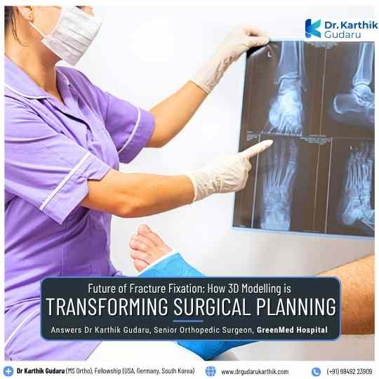
Conventional X-rays provide important information about a fracture, while CT scans can offer detailed cross-sectional views of the affected bone. These CT images can also be processed to create three-dimensional models, allowing surgeons to study the anatomy and fracture pattern from different angles.
This additional perspective can be particularly valuable when a fracture is complex or when the normal anatomy is difficult to visualize from conventional imaging alone.
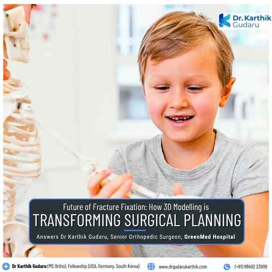
“3D-printed models can give surgeons a clearer understanding of complex fractures and help them visualize the bone anatomy before planning fixation.” says Dr Gudaru Karthik.
Better Understanding of Complex Fractures
Some fractures are difficult to fully understand from standard two-dimensional images, particularly when the bone is displaced, rotated or fractured into multiple pieces.
A three-dimensional model can provide a more realistic representation of the fracture, helping the surgical team visualize the location and orientation of the fracture fragments.
This can be especially useful when planning the treatment of complex fractures where understanding the three-dimensional anatomy is important.
More Precise Fracture Fixation Planning
Before surgery, surgeons can use 3D models to study the fracture and consider different approaches to fixation. A physical model may also help them visualize where reduction, drilling, cutting or implant placement may be required.
This preparation allows the surgical plan to be tailored to the patient’s specific anatomy rather than relying entirely on standard approaches.
The goal is not simply to use advanced technology, but to use it when it can provide meaningful information that supports better surgical planning.
Understanding the Patient’s Unique Anatomy
Every patient’s bone anatomy is different. In complex fractures, previous injuries, deformities or anatomical variations can make it difficult to rely on standard assumptions about bone shape and alignment.
A 3D model can help surgeons appreciate these individual differences before surgery.
The ability to compare the fractured anatomy with the patient’s normal, non-fractured side in selected cases may provide an additional reference when determining the original shape and alignment of the bone.
Potential Role of Patient-Specific Surgical Guides
The development of 3D technology has also opened possibilities for patient-specific surgical guides.
These guides can be designed using the patient’s imaging data and may help surgeons translate a carefully planned procedure into the operating room.
Their usefulness depends on the type of fracture, the complexity of the procedure, available technology and the surgeon’s clinical judgment. They are not required for every fracture, but may have a role in selected complex cases.
Can 3D Models Improve Surgical Communication?
Medical images can sometimes be difficult for patients and families to interpret. A physical three-dimensional model may provide a more intuitive way to demonstrate the location and nature of a fracture.
This can help patients better understand their condition and have more informed discussions with their surgeon about the proposed fixation procedure.
Technology Supports the Surgeon—It Does Not Replace Expertise
3D modeling is a planning and visualization tool, not a replacement for surgical experience or clinical judgment.
The surgeon still needs to consider the patient’s symptoms, examination findings, imaging, bone quality, fracture characteristics, overall health and expected recovery before deciding on treatment.
Technology is most valuable when it complements the surgeon’s expertise and provides additional information that can contribute to thoughtful surgical planning.
What Could the Future of Fracture Fixation Hold?
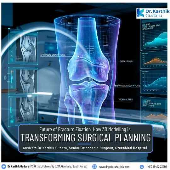
As medical imaging, computer-assisted planning and 3D printing technologies continue to develop, their role in fracture fixation may expand.
Future applications could include increasingly personalized surgical planning, improved preoperative simulation and more patient-specific solutions for complex fractures.
However, the value of any technology ultimately depends on whether it can meaningfully improve surgical planning, patient safety and clinical outcomes.
3D modeling is therefore not about replacing established orthopaedic techniques. Instead, it represents another step towards more personalized fracture fixation, where surgeons can understand the patient’s anatomy in greater detail and prepare for complex procedures before entering the operating room.

Dr Karthik Gudaru, MS Orthopaedics, MBBS, Fellowship in Knee and Hip Arthroplasty (USA), Fellowship in Computer-Assisted Navigation Joint Replacement Surgery (Germany), is one of Hyderabad’s leading orthopaedic surgeons with an outstanding reputation for precision, innovation, and excellence in joint replacement and orthopaedic care. Widely recognised as the best knee replacement specialist in Hyderabad, he brings a deep commitment to patient-centred care and cutting-edge surgical techniques.
Dr Karthik Gudaru is currently the Senior Orthopaedic and Joint Replacement Specialist, Department of Orthopaedics at GreenMed Hospital, Srinagar Colony, Hyderabad; Assistant Professor and Consultant Orthopaedic at BIRRD Hospital (Balaji Institute of Surgery, Research and Rehabilitation for the Disabled), Tirupati; and Joint Replacement & Pediatric Orthopaedic Specialist at Sri Maruthi Hospitals, Tirupati.
Dr Karthik Gudaru’s treatment specialisations –
Total Knee Replacement Surgery.
Total Shoulder Replacement Surgery.
Hip Joint Treatments
Total Elbow Arthroscopy.
Computer-Assisted Knee Replacement Surgery.
Stem Cell Therapy for key orthopaedic treatments.
Pediatric Orthopaedics Treatments for Cerebral Palsy, Congenital Anomalies, Poliomyelitis & Metabolic Bone Diseases
Hospital Affiliations
OPD, GreenMed Hospital, 8-3-833/71, Srinagar Colony Main Rd, Kamalapuri Colony, Banjara Hills, Hyderabad, Telangana
Durgabai Deshmukh Hospital, Vidyanagar, Hyderabad
Sri Maruthi Hospitals, OPD, 19-3-30/C1, Near RTC Bus Stand, Kennedy Nagar, Tirupati, 517501
BIRRD Hospital, Tirupati (Consultant & Asst. Professor)
Education and Affiliation
MBBS: Kasturba Medical College, Manipal University, Karnataka (2012), Registration No: TSMC/FMR/16001
MS (Orthopaedics): Sri Ramachandra Medical College & Research Institute, Chennai (2016), Registration No: 119525
Knee & Hip Arthroplasty Fellowship: Dartmouth Hospital, USA (2018)
Robotic & Computer-Assisted Joint Replacement Fellowship: Asklepios Hospital, Germany (2018)
AO Trauma & Emergency Orthopaedic Care Fellowship: Guro National Hospital, South Korea (2019–2020)
Spinal Surgery Observer Training: Yonsei University, South Korea (2015)